

Cellular Analysis
We offer support in light microscopy (live-cell imaging, confocal, super-resolution, intravital, high-throughput), electron microscopy (transmission and scanning electron microscopy, immunolabeling, cryo-microscopy), in vivo imaging, Histology and Flow Cytometry; as well as Image Analysis support.
We provide training on our systems, as well as full support from sample handling to imaging/data collection and data analysis.
Electron Microscopy
We can provide a high-quality service for students and staff across the University, enabling state-of-the-art preparation of a wide range of biological and material specimens.We also welcome researchers from other academic institutions and commercial organisations.
Support and advice is provided for consultation prior the work and throughout the sample processing and imaging by technical staff with several years of experience.
What we offer:
- Transmission Electron Microscopy
- Scanning Electron Microscopy
- Specialised Cryo-preparations Techniques
- Immunolabelling
- Electron Tomography
Learn more about Electron Microscopy and the equipment we use:
Our Equipment
- Leica EM UC7 with EM FC7 cryochamber
- JEOL IT 100 SEM
- JEOL 1400Flash TEM
- Leica EM UCT
- Reichert Ultracut E
- Reichert Knifemaker
- Leica EM GP2
- Tousimis autosamdri-815 Critical point dryer
- Quorum Q150T ES multifunction high vacuum coater
- Eppendorf Centrifuge 5804 R with temperature control
Transmission Electron Microscopy
- Conventional resin embedding of biological specimens
- Semi-thin and Ultra-thin sectioning for light and TEM microscopy
- Low temperature specimen processing for Immunocytochemistry
- Negative staining and Methylcellulose/Uranyl Acetate embedding
Scanning Electron Microscopy
- Critical point drying
- High vacuum metal sputter coating
Specialized Cryo-preparations techniques
- Cryo-Ultramicrotomy
- Freeze Drying
- Freeze Fracture
Flow Cytometry
The Flow Core Facility provides access to a range of Flow Cytometers for the whole University community.
Our machines can be used for multicolour immunophenotyping, cell cycle analysis, identification and isolation of fluorescent protein transfected cells, isolation of rare cell populations, analysis of bacteria and parasite populations, apoptosis assays, and functional assays.
What we offer:
- Cell Sorting
- FACS Analysis
- Spectral FACS Analysis
Learn more about our Flow Cytometry equipment:
Curiox HT2000
Our Curiox Laminar Wash HT2000 System can be used for sample preparation for flow cytometry and for omics applications. It is particularly useful for fragile cells as the gentle laminar washing procedure causes less mechanical stress on cells and less cell loss compared to conventional centrifugation.
Advantages of the system are:
- Reduce the amount of aerosolisation compared to a centrifugation
- Increase Cell Retention
- Gives higher percentages of viable cells after processing compared to centrifugation.
- Speed – The system completely washes 96 samples in 4 minutes
- Reduced background with more thorough washing.
- No Pelleting of Cells – reducing doublets and clumping.
- Higher Stain Index – For better resolution of populations
- Cleaner Data – Improved cell segregation and resolution; Reduces debris and aggregation of cells
Curiox plates and large volume adapters areavailable at cost price from Flow Core staff.
We offer instrument training, and assistance with adapting staining protocols for the curiox.
BD LSR Fortessa Analyser
We have a SORP BD LSR Fortessa flow cytometer. This machine has five lasers: Blue (488mn), Red (640nm), Violet (405nm), Yellow Green (561nm) and UltraViolet (305nm). It can detect 20 parameters simultaneously (18 colours plus forward scatter and side scatter).
Digital pulse sampling allows the measurement of Height, Area and Width from all parameters simultaneously.
Data is acquired through BD FACSDiVaTM digital software.
Typical operation at 20,000 events/ second which requires a sample concentration of 2 x 10⁷ cells/mL at an instrument flow rate of 60 μL/min.
The LSR Fortessa also has a High Throughput Option for analysis. This provides rapid, fully automated sample acquisition from microtiter plates. The HTS option supports a wide range of research applications and is compatible with 96-well U, V, and flat-bottom plates as well as 384-well microtiter plates. In high-throughput mode, the HTS can process a 96-well plate in fewer than 15 minutes with less than 1 per cent carryover. Standard throughput mode can be selected to acquire larger sample volumes or where longer acquisition times are required.
The beauty of this machine is that filter and mirror changes can easily be made to customise the optical array for non standard experiments.
For access to this machine please contact the Flow Core Facility Manager.
Applications: Analysis of cells, parasites, bacteria, calcium flux, phospho flow, DNA analysis/cell cycle, apoptosis and multi colour immunophenotyping, Fluorescent proteins, cell proliferation etc.,
BD Celesta Analyser
The newest addition to the lab is a BD Celesta flow cytometer.
This machine is equipped with 3 lasers and can detect 14 parameters simultaneously.
The BD FACSCelesta is the first flow cytometer optimized by design to work with BD Horizon Brilliant dyes The Celesta fits neatly on our benchtop.
Our Celesta has a HTS installed to improve experimental workflow, the HTS provides rapid, fully automated sample acquisition from 96- and 384-well microtiter plates.
For most applications, standard throughput mode is optimal and can be selected for acquisition of larger sample volumes. For selected applications, the high-throughput option, can speed through a 96-well plate in fewer than 15 minutes with less than 0.5% sample carryover from one well to the next.
For access to this machine please contact the Flow Core Facility Manager.
Applications: Analysis of cells, parasites, bacteria, DNA analysis/cell cycle, apoptosis and multi-colour immunophenotyping, Fluorescent proteins, cell proliferation, rare cell detection etc.,
BD Aria Cell Sorter
The Facility offers a cell sorting service.
We have a BD FACSAria IIU, and a BD FACSAria III high speed cell sorter. These provide sterile, high purity multi colour sorts.
Both instrument configurations are identical, with four lasers each: Violet (405nm),Blue (488mn), Red (640nm) and Yellow Green (561nm) and 18 fluorescence parameters.
The machines sort a range of particle sizes into a maximum of 4 separate populations, into microtitre or cell culture plates or into tubes. Cells can be sorted in a sterile environment enabling the recovered cells to be cultured. This allows selective sorting for cloning, or enrichment of populations. We also sort to purify cell populations prior to PCR amplification, or for protein analysis. Filter and mirror changes can easily be made to customise the optical array for non standard experiments.
Data is acquired through BD FACSDiVaTM digital software.
To book a Sort please contact the Flow Core Facility Manager to discuss your requirements. Time on the sorter is at a premium. You are advised to make a booking several weeks in advance of your planned sort date.
Applications : Sorting of cells, parasites, etc based on multi colour phenotypic markers, DNA /cell cycle analysis, DNA dye exclusion (side populations), expression of fluorescent proteins, clonal sorts ,Index sorting, Single cell sorting for RNA seq etc.,
BD Canto II
New to the Facility as of September 2021 is Clyde, the BDFACS Canto II flow cytometer.
This machine has three lasers: Blue (488mn), Red (640nm) and Violet (405nm). It can detect 10 parameters simultaneously (8 colours plus forward scatter and side scatter).
Digital pulse sampling allows the measurement of height, area and width from all parameters simultaneously.
Data is acquired through BD FACSDiVaTM digital software.
Typical operation at 20,000 events/ second which requires a sample concentration of 2 x 10⁷ cells/mL at an instrument flow rate of 60 μL/min.
The beauty of this machine is that filter and mirror changes can easily be made to customise the optical array for non standard experiments. Additionally we are able to switch off selected lasers as required for specialist applications.
Sony ID7000 Spectral Analyser
Our newest analyser, the Sony ID7000 is a 5-laser instrument (355nm, 405nm, 488nm, 561nm, 660nm) with 147 detectors. It is different to our conventional cytometers as it doesn’t use physical filter sets (so we don’t have a traditional configuration document) but rather it takes all of the light emitted and unmixes the spectral signatures of individual fluorochromes.
Whilst this is a different way of working the instrument has intuitive software for experiment setup, spectral unmixing and autofluorescence extraction.
The analysis software is free to all users of the system but runs on a PC and requires 16GB of RAM.
Acquired data must be unmixed at the instrument, but thereafter can be analysed off line.
As we look in more detail at our cells, the number of parameters needed increases, and with the proper advice and experimental design, panels of over 40 colours are now possible.
The instrument has an autosampler and walk away analysis is truly possible once settings have been optimised.
Sony MA900 Cell Sorter
Our Sony MA900 cell Sorter meets the needs of most cell sorting applications, with 12 fluorescence parameters and up to 4-way sorting into tubes, or 96 and 884 well plate sorting.
The MA900 uses a patented disposable micro fluidic chip base design and has automated sorter setup with troubleshooting wizards that simplify the operation of the instrument. It is situated in a Class 2 Biosafety cabinet.
Sorting chips are included for cost price from the flow core staff, as are sample lines if you wish to use your own every time you sort. We also have unlimited free copies of the analysis version of the sorter software, which we will share with users of the instrument. The software runs on PC’s. Please contact us for training or advice.
The hourly cost for this is the same as the BD FACS Aria sorters, however a charge of £20 is added to each booking to cover the cost of the disposable chip.
BD FAC Symphony Cell Analyser
This is our second instrument with spectral capabilities. It is a 5 laser system, measuring up to 48 parameters.
This features an optimised filter set to collect the full spectrum of emitted light, increasing flexibility in fluorochrome choices and enabling simultaneous analysis of fluorochromes with similar spectral signatures.
Using the same BD FACSDiva™software already on our existing conventional analysers, allows users to confidently move to the world of spectral flow cytometry.
Histology
The Histology Core Facility can provide routine histology services for researchers within the School of Immunology and Infection and throughout the wider University. We are a CL2 lab meaning we can handle both mouse and human tissue as well as various infectious materials. Services we offer include tissue processing, embedding and sectioning for formalin-fixed tissue and frozen tissue. We provide H&E staining, special staining, immunohistochemistry and immunofluorescence. We also provide brightfield slide scanning services to image whole-tissue sections.
What we offer:
- Tissue Processing and Embedding
- Microtomy
- Cryosectioning (cryotome)
- Special Staining
- Immunohistochemistry and Immunofluorescence
- Slide Scanning
Learn more about our Histology equipment:
Tissue Processing and Embedding
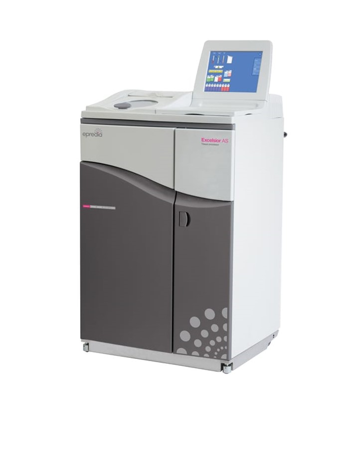
The facility has an Epredia Excelsior AS processor, capable of processing up to 300 cassettes in a single run. You can input and save multiple processing programmes and the system monitors reagent quality ensuring optimal processing. Samples should be provided in 70% Ethanol, already grossed and ready to be transferred into a cassette. Following processing, we can embed the samples into paraffin for sectioning and further staining.

Microtomy
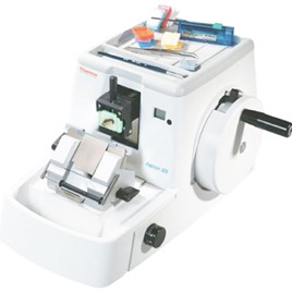
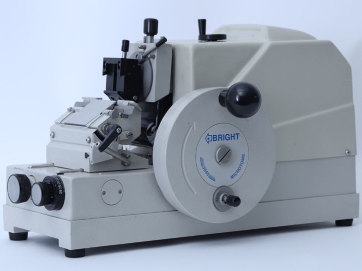
We have two manual rotary microtomes used to section FFPE tissue blocks, the Shandon Finesse 325 and the Bright 5040 both have adjustable section thickness, clearance angle and block holder.
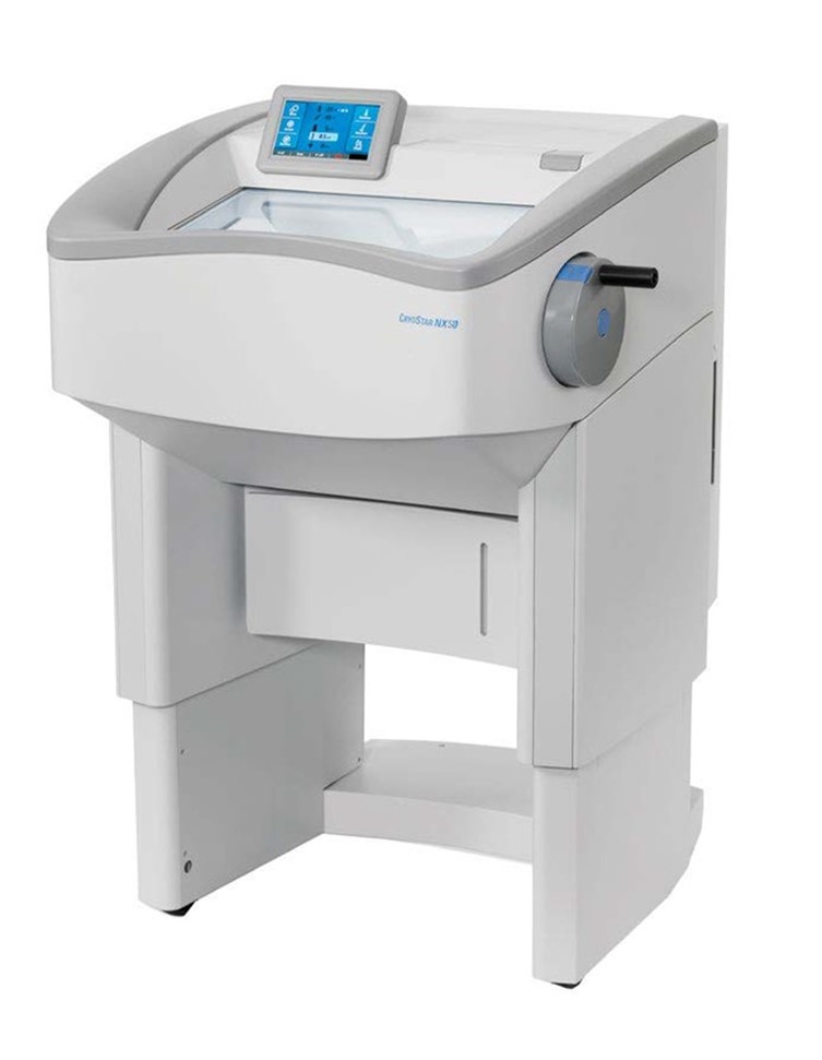
Cryotomy
We offer sectioning of fixed-frozen or fresh-frozen tissue, using the Epredia NX50 Cryostat which features cooling of the specimen head, blade holder and chamber, with rapid temperature adjustments. The system also features motorised height control to allow comfortable working.
Special Staining
We can provide H&E staining as well as special staining, including any optimisation of stains. Please email the Histology Core Facility Manager to discuss your specific requirements further.
Immunohistochemistry and Immunofluorescence
We provide a service for immunohistochemistry and immunofluorescence of tissue sections and have a stock of commonly used antibodies available for use. We can also optimise and stain with antibodies purchased by the user and offer advice on protocol development.
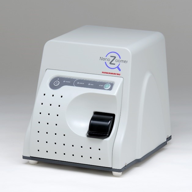
Slide Scanning
We offer high-speed, brightfield slide scanning, using the Hamamatsu Nanozoomer-SQ. This features 20X or 40X scanning in a single Z-plane or as a Z-stack. The system is user-friendly and produces high quality scans of large tissue sections.
*** We also provide training in all these techniques with the exception of tissue processing, where training will only be provided if routine use is needed.
Light Microscopy
The Light Microscopy Core has imaging technologies that cover whole body imaging (resolution in mm), through conventional wide-field, confocal and multi-photon microscopy (resolution in µm), to high-resolution approaches breaking the resolution limit of light (resolution in nm).
What we offer:
- Confocal Microscopy
- Live-cell confocal
- Spinning Disk Confocal
- Multiphoton
- High-Throughput Content
- Super-Resolution (3D-SIM and STORM)
- Live-cell wide-field microscopy with deconvolution
- Imaging flow cytometry